
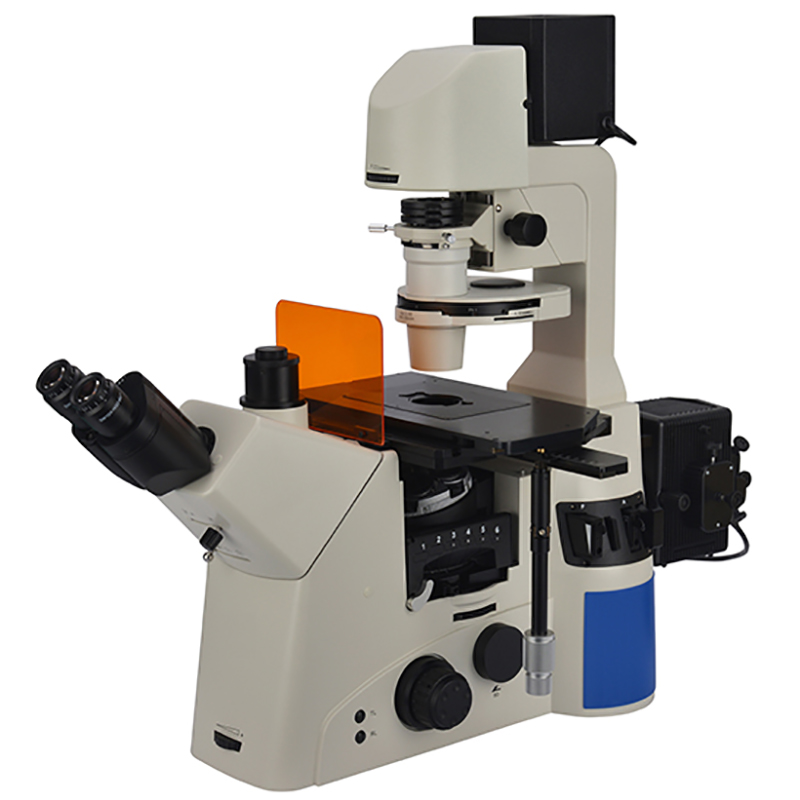
Fluorescence microscopy has revolutionized our ability to visualize and study biological specimens, allowing us to delve into the intricate world of cells and molecules. A key component of fluorescence microscopy is the light source used to excite fluorescent molecules within the sample. Over the years, various light sources have been employed, each with its unique characteristics and advantages.
1. Mercury Lamp
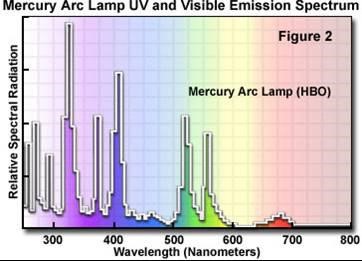
The high-pressure mercury lamp, ranging from 50 to 200 watts, is constructed using quartz glass and is spherical in shape. It contains a certain amount of mercury inside. When it operates, a discharge occurs between two electrodes, causing mercury to evaporate, and the internal pressure in the sphere rapidly increases. This process typically takes about 5 to 15 minutes.
The emission of the high-pressure mercury lamp results from the disintegration and reduction of mercury molecules during the electrode discharge, leading to the emission of light photons.
It emits strong ultraviolet and blue-violet light, making it suitable for exciting various fluorescent materials, which is why it is widely used in fluorescence microscopy.
2. Xenon Lamps
Another commonly used white light source in fluorescence microscopy is the xenon lamp. Xenon lamps, like mercury lamps, provide a broad spectrum of wavelengths from ultraviolet to near-infrared. However, they differ in their excitation spectra.
Mercury lamps concentrate their emission in the near-ultraviolet, blue, and green regions, which ensures the generation of bright fluorescent signals but comes with strong phototoxicity. Consequently, HBO lamps are typically reserved for fixed samples or weak fluorescence imaging. In contrast, xenon lamp sources have a smoother excitation profile, allowing for intensity comparisons at different wavelengths. This characteristic is advantageous for applications like calcium ion concentration measurements. Xenon lamps also exhibit strong excitation in the near-infrared range, particularly around 800-1000 nm.
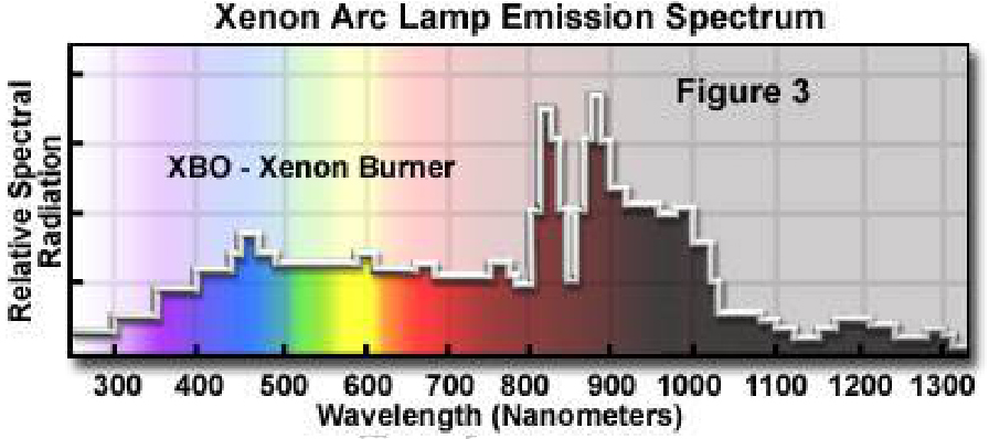
XBO lamps have the following advantages over HBO lamps:
① More uniform spectral intensity
② Stronger spectral intensity in the infrared and mid-infrared regions
③ Greater energy output, making it easier to reach the objective's aperture.
3. LEDs
In recent years, a new contender has emerged in the realm of fluorescence microscopy light sources: LEDs. LEDs offer the advantage of rapid on-off switching in milliseconds, reducing sample exposure times and extending the lifespan of delicate samples. Furthermore, LED light exhibits quick and precise decay, significantly diminishing phototoxicity during long-term live cell experiments.
Compared to white light sources, LEDs typically emit within a narrower excitation spectrum. However, multiple LED bands are available, allowing for versatile multi-color fluorescence applications, making LEDs an increasingly popular choice in modern fluorescence microscopy setups.
4. Lasers Light Source
Laser light sources are highly monochromatic and directional, making them ideal for high-resolution microscopy, including super-resolution techniques such as STED (Stimulated Emission Depletion) and PALM (Photoactivated Localization Microscopy). Laser light is typically selected to match the specific excitation wavelength required for the target fluorophore, providing high selectivity and precision in fluorescence excitation.

The choice of a fluorescence microscope light source depends on the specific experimental requirements and sample characteristics. Please feel free to contact us if you need any help
Post time: Sep-13-2023