
There are more and more types of microscopes, and the scope of observation is also wider and wider. Roughly speaking, they can be divided into optical microscopes and electron microscopes. The former uses visible light as the light source, and the latter uses electron beams as the light source. Optical microscopes can be divided into different types according to their structure, observation method and use.
In this article, we will divide them into the 9 most common types according to their use, so that you can better understand the microscope and choose the right product.
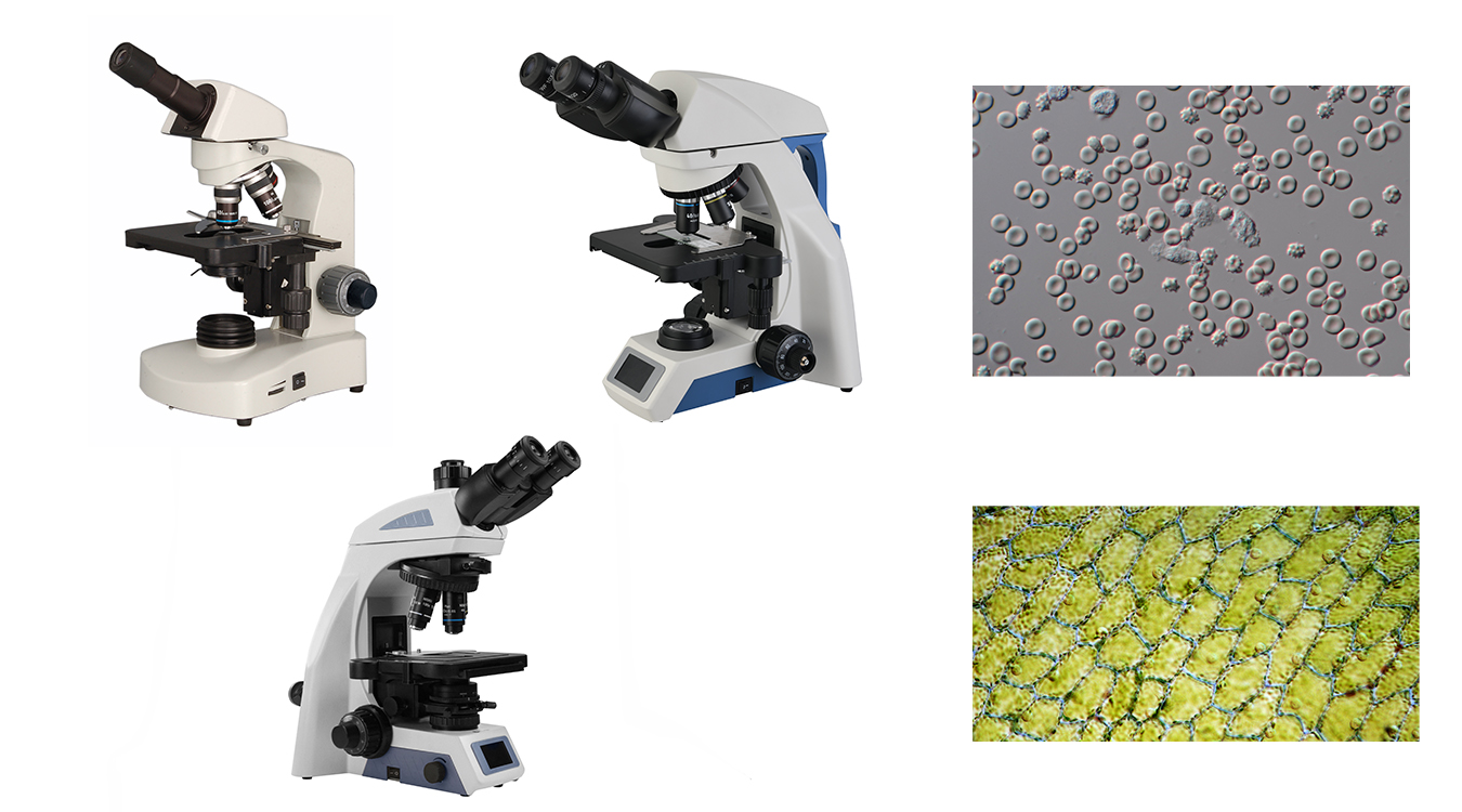
- Biological Microscope
The optical part of a biological microscope includes eyepieces and objective lenses. The objective lens is the core component of the microscope. The most common objectives are 4x, 10x, 40x, and 100x, which are divided into three levels: achromatic, semi-plan achromatic, and plan achromatic. Optical systems can be divided into finite objectives and infinite objectives. Plan achromatic objectives have no defects in the field of view and are commonly used in scientific research and medical specialties. Microscope head can be divided into monocular, binocular and trinocular head. Binocular microscopes can see samples with two eyes at the same time. Additional eyepieces for trinocular microscope can be attached to cameras or digital eyepieces to display images, measure and analyze as needed for work or research.
Commonly viewed samples include biological slides, biological cells, bacteria and tissue culture, liquid sedimentation. Biological microscopes can be used for observation, diagnosis and research of sperm, blood, urine, feces, tumor cell pathology and so on. Biological microscopes also can be used to observe transparent or translucent objects, powders and fine particles, etc.
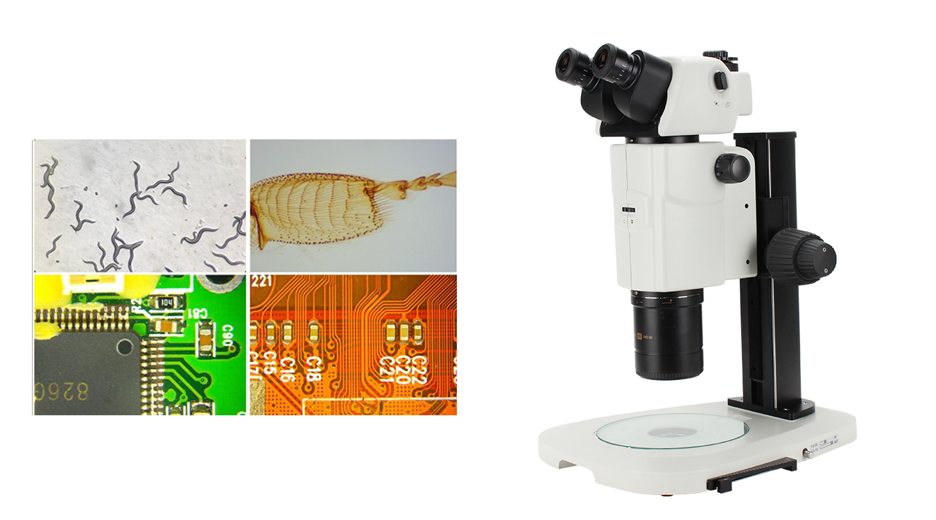
- Stereo Microscope
Stereo microscopes work by using two light paths at slightly different angles to produce a three-dimensional view of the sample under the lens, which can be observed through the binocular eyepieces. Typically, 10x to 40x magnification is available, and this lower magnification, coupled with a larger field of view and working distance, allows more manipulation of the object under observation. For opaque objects, it uses reflected lighting for better 3D viewing.
Stereo microscopes are commonly used in the manufacture of items such as circuit boards, electronics, semiconductor and botanical observation and study. Stereo microscopes also can be used for various experiments and research like animal anatomy teaching, test tube babies and life sciences.
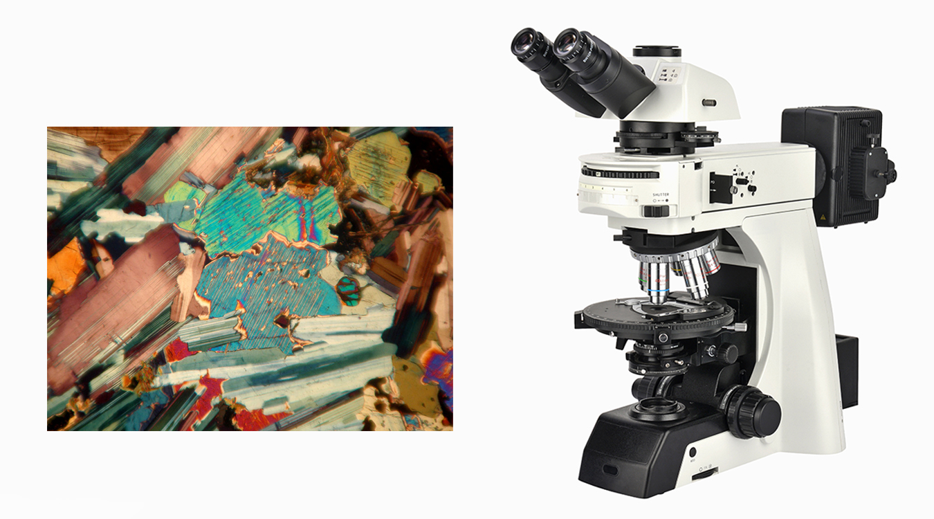
Polarizing Microscope
Polarizing Microscopes use light manipulation to increase the contrast between different structures and densities under magnification. They use transmitted and/or reflected light, filtered by a polarizer and controlled by an analyzer, to highlight differences in texture, density, and color on the sample surface. Therefore, they are ideal for viewing birefringent materials.
Polarizing microscopes are often used in geology, petrology, chemistry and many other similar industries.
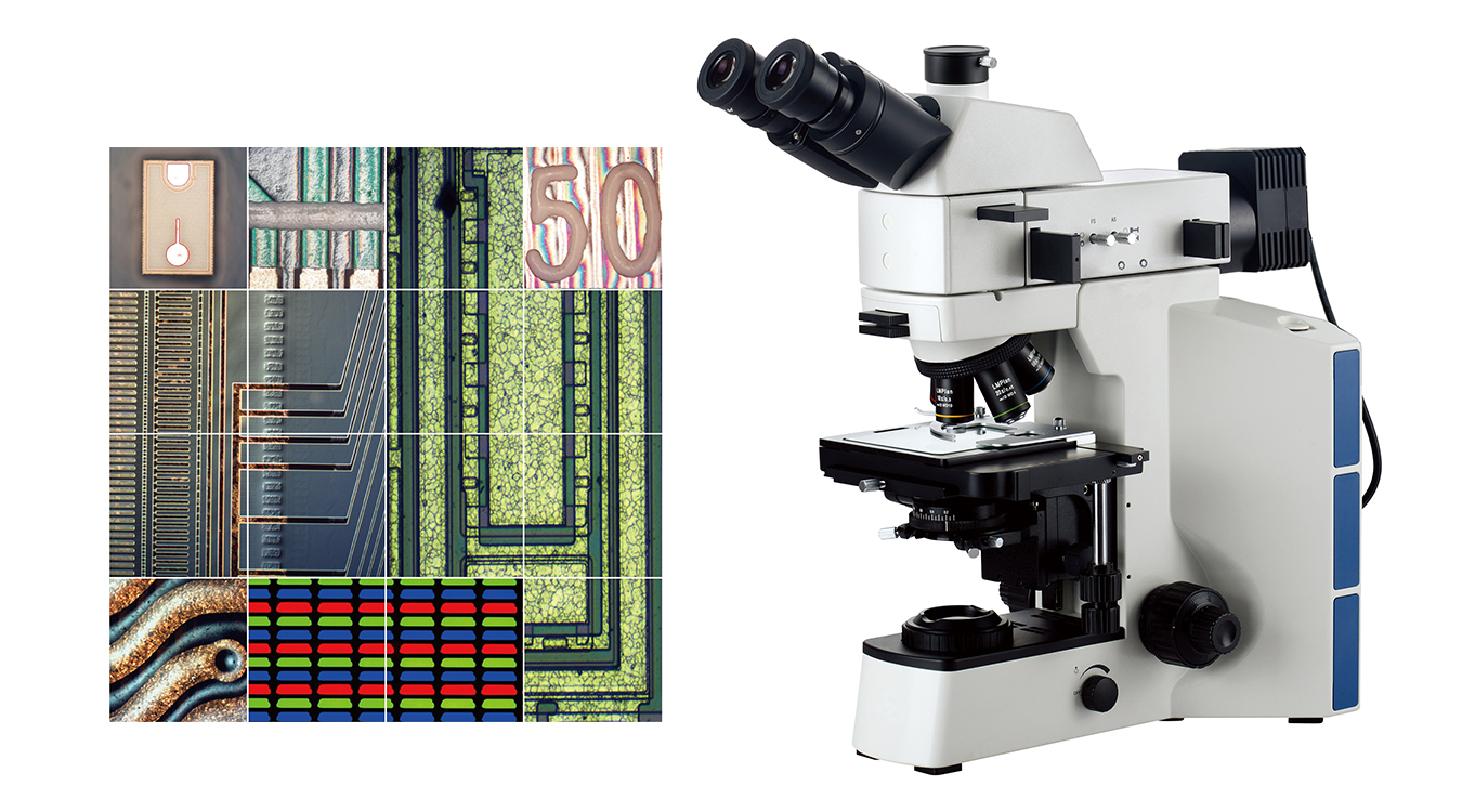
Metallurgical Microscope
Metallurgical microscopes are high-powered microscopes designed to observe samples that do not allow light to pass through. The reflected light shines through the objective lens, providing magnifications of 50x, 100x, 200x, 500x, and sometimes even 1000x. Metallographic microscope is used to examine microstructure, micron-scale cracks, very thin coatings such as paint and grain size in metals.
Metallographic microscopes are used in the aerospace industry, automotive manufacturing, and companies that analyze metal structures, composites, glass, wood, ceramics, polymers, and liquid crystals. They can also be utilized for related products in the semiconductor industry and the inspection and analysis of wafers.
Fluorescent Microscope
Fluorescent Microscopes emit light onto cells stained with fluorescent dyes, allowing cell features to be seen more clearly than conventional microscope using reflected light. Fluorescent Microscopes are also highly sensitive and can detect differences in brightness and wavelength. This makes it possible to observe details that cannot be seen with standard white light optical microscopes.
It is commonly used in biology and medicine to study cellular proteins and identify bacteria in living organisms.

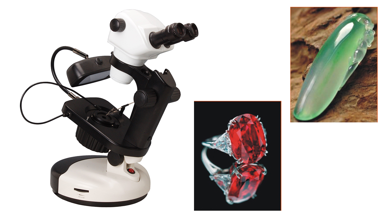
Gemological Microscope
Gemological Microscope is a vertical double simple stereo continuous zoom microscope. The commonly used magnification is 10 to 80 times. It is equipped with a bottom light source and a top light source, it is also equipped with a dark field illumination used with the bottom light source, adjustable diaphragm and gemstone clips. It allows users to conduct multi aspect observation and research on gemstones using transmitted or reflected methods.
It is used to observe and evaluate gemstones of different types and grades, as well as gemstone setting, assembly, and repair.
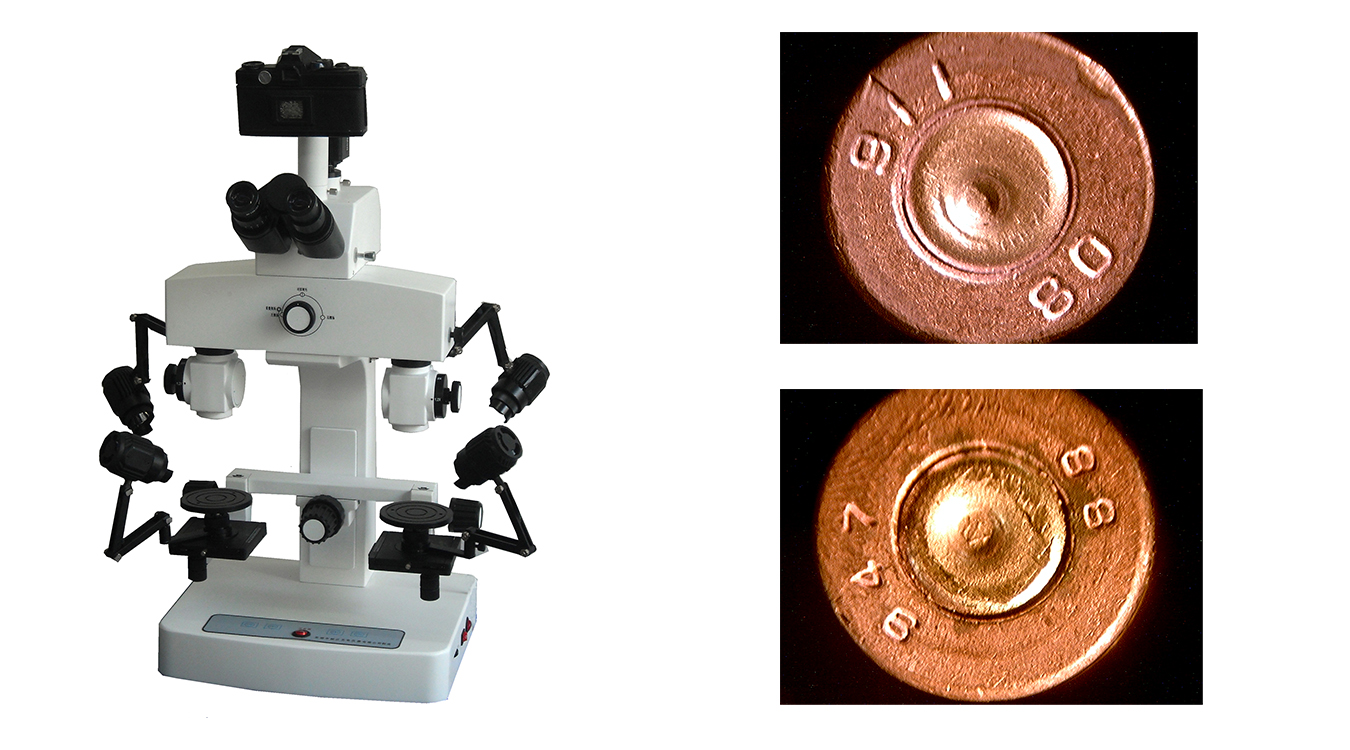
Comparison Microscope
Comparison microscopes are special microscopes, they are also called forensic microscopes. It not only has the magnification effect of ordinary microscope, but also can observe the image of the object left and right in optical systems simultaneously with a set of eyepieces. It can compare two or more objects macroscopically or microscopically to examine, analyze and identify their minor differences in form, organization, structure, color or material through docking, cutting, overlapping, rotating, etc. In order to achieve the purpose of identification and comparison.
The main application of these types of dual microscopes are in criminology and ballistics. They are the mainstay of forensic science, too. Other scientific fields, including paleontology and archaeology, also use these special compound microscopes.
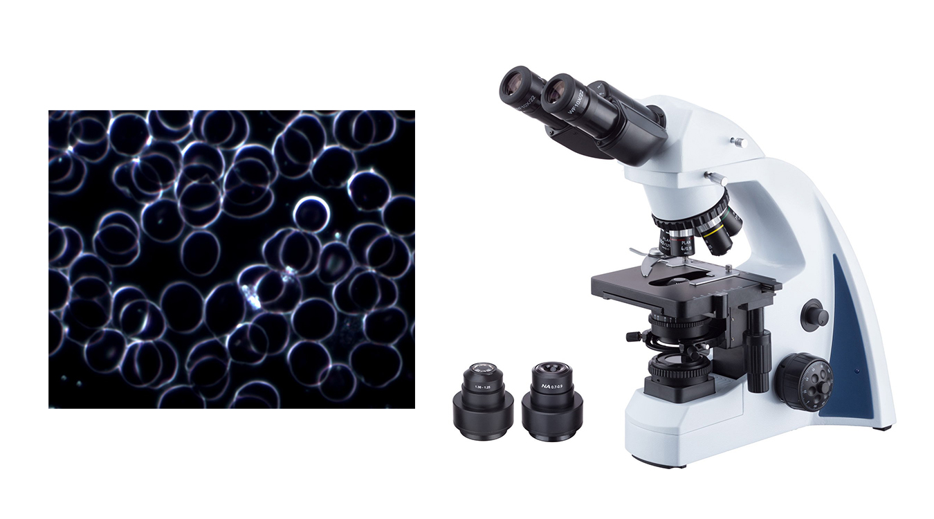
Dark Field Microscope
There is a light sheet in the center of the condenser of a darkfield microscope, so that the illumination light does not directly enter the objective lens, and only the light reflected and diffracted by the specimen is allowed to enter the objective lens, so the background of the field of view is black, and the edge of the object is bright. Using this microscope, microparticles as small as 4-200 nm can be seen, and the resolution can be 50 times higher than that of ordinary microscopes.
Darkfield illumination is particularly suitable for showing contours, edges, boundaries and refractive index gradients. For observation of tiny aquatic organisms, diatoms, small insects, bone, fibers, hair, unstained bacteria, yeast, tissue culture cells and protozoa.
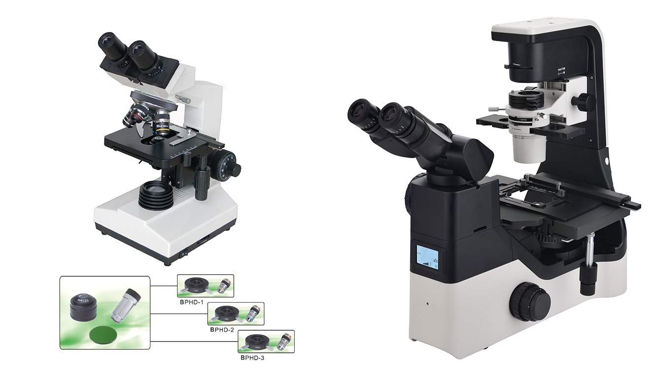
Phase Contrast Microscope
Phase contrast microscope uses the diffraction and interference phenomena of light to convert the optical path difference or phase difference of the light passing through the specimen into an amplitude difference microscope that can be resolved by the naked eye. The difference between light and dark in images of substances with different densities is improved, which can be used to observe unstained cell structures. Phase contrast microscopes can be divided into upright phase contrast microscopes and inverted phase contrast microscopes.
It is mainly used for the cultivation and observation of sperm, living cells and bacteria, as well as providing special functions such as observation of embryo morphology and differentiation of embryo stages.
Hope the above content can help you choose the right microscope type, if you have any questions, please contact us.
Post time: Sep-06-2022