
The bright field observation method and the dark field observation method are two common microscopy techniques, which have different applications and advantages in different types of sample observation. The following is a detailed explanation of the two methods of observation.
Bright Field Observation Method:
The bright field observation method is one of the most fundamental and widely used microscopy techniques. In bright field observation, the sample is illuminated with transmitted light, and the image is formed based on the intensity of the transmitted light. This method is suitable for many routine biological specimens, such as stained tissue slices or cells.
Advantages:
Easy to operate and applicable to a wide range of biological and inorganic samples.
Provides a clear view of the overall structure of biological specimens.
Disadvantages:
Not suitable for transparent and colorless samples, as they often lack contrast, making it challenging to obtain clear images.
Unable to reveal fine internal structures within cells.
Dark Field Observation Method:
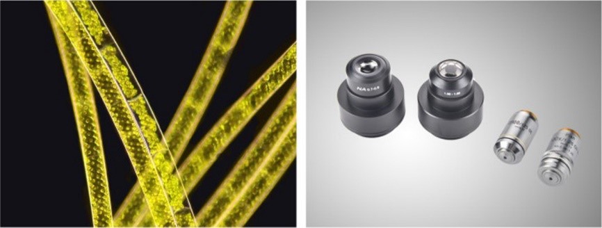
Dark field observation utilizes a specialized lighting arrangement to create a dark background around the sample. This causes the sample to scatter or reflect light, resulting in a bright image against the dark background. This method is particularly suitable for transparent and colorless samples, as it enhances the edges and contours of the sample, thereby increasing contrast.
A special accessory required for dark field observation is a dark field condenser. It is characterized by not letting the light beam pass the object under inspection from the bottom up, but changing the path of the light so that it is slanted toward the object under inspection, so that the lighting light does not directly enter the objective lens, and the bright image formed by the reflection or diffraction light on the surface of the object under inspection is used. The resolution of dark field observation is much higher than that of bright field observation, up to 0.02-0.004μm.
Advantages:
Applicable for observing transparent and colorless samples, such as live cells.
Enhances the edges and fine structures of the sample, thereby increasing contrast.
Disadvantages:
Requires a more complex setup and specific equipment.
Involves adjusting the positioning of the sample and light source for optimal results.
Post time: Aug-24-2023